New Imaging Techniques Spot HCM Before it Develops

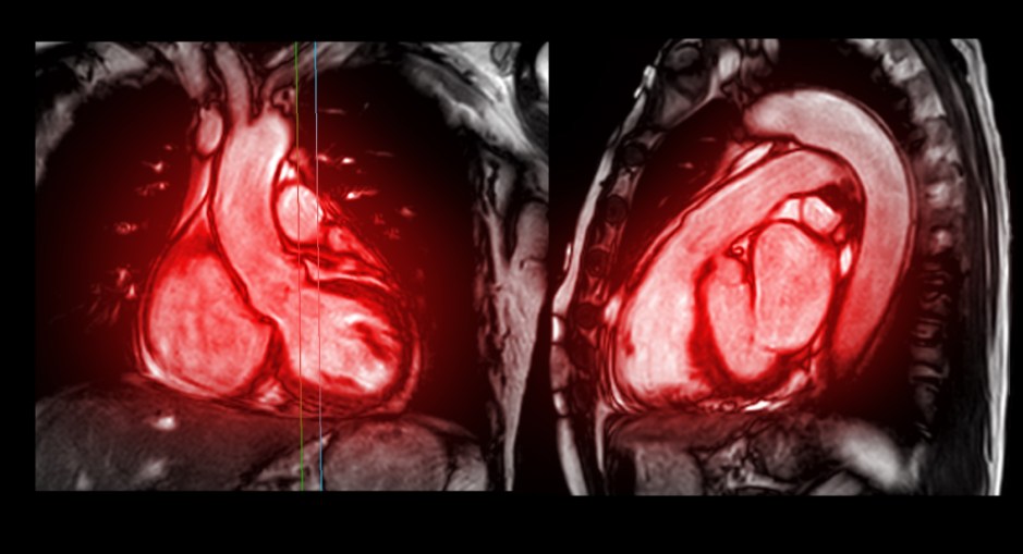
Research funded by the British Heart Foundation using novel cardiac imaging techniques was described in a recent article in Circulation. These techniques, such as quantitative perfusion cardiac magnetic resonance imaging and cardiac diffusion tensor imaging, can detect pre-clinical hypertrophic cardiomyopathy (HCM) before the development of any thickening in the heart.

With novel treatments like myosin modulators now available to patients, and with  experimental treatments such as gene therapy now on the horizon, these new imaging techniques could allow doctors to intervene at a much earlier stage in order to potentially prevent disease.

You can read more about these imaging techniques here.

New Technology May Find Early Signs of HCM

A recent paper published in Nature Reviews Cardiology shows how new advances in Cardiac MRI and biomarkers identified through blood testing may help to identify subclinical HCM before overt signs are evident.

This information could be used to intervene before patients ever develop overt signs of disease. Newer medications or experimental gene therapies have the potential to prevent the condition from ever developing.

These modalities would be useful when screening  relatives of HCM patients and/or in people known to carry a gene for HCM.

New MRI Technology Spots Scar Tissue Using AI

A new technology called “virtual native enhancement” (VNE) may soon eliminate the need for gadolinium as a contrast agent for patients with hypertrophic cardiomyopathy undergoing cardiac MRI. Gadolinium, a heavy metal contrast agent which is injected intravenously, has long been used in cardiac MRI scans to spot cardiac scar tissue in patients with HCM.  In 2017, the FDA issued a safety communication relating to gadolinium  because it was found that gadolinium remains in the body for months to years after the use of the drug.

The new VNE technology, recently described in the journal Circulation, uses artificial intelligence (AI) to virtually enhance the standard MRI image. The technology was developed using data taken from 1348 HCM patients and was validated in the HCM population, but the technology may have uses extending beyond HCM.

By avoiding the use of the contrast agent, this technology avoids side effects and long term consequences from the use of gadolinium. Additionally, it will make cardiac MRI available to patients who are allergic to gadolinium. VNE is also faster and cheaper that current technology used for cardiac MRI, which may make more frequent MRI monitoring of patients feasible.  

To read more about VNE, see also this article in UVA Today, this article in ACM Tech News,  this article in Engineering and Technology, and this article in Science Daily.

Can Cardiac MRI Predict AFib in HCM Patients?

In a recent study, researchers examined whether cardiac MRI results might help predict which patients would would go on to develop atrial fibrillation (AFib) that was serious enough to require hospitalization, require electrical cardioversion or catheter ablation, or identify those patients who might go on to develop permanent AFib.

The study found that the major predictors of these serious AFib consequences in HCM were those who were of older age, those with an increased BMI (this was especially important in patients under age 33), increased left atrial volume index as seen on cardiac MRI (this was especially important in middle-aged patients), reduced left atrial contractile function (this was especially important in middle-age and older patients), and moderate or severe mitral valve regurgitation. 

The researchers concluded that using cardiac MRI to measure left atrial volume and contractile function might help medical providers ability to intervene before major AFib develops in HCM patients, specifically by helping patients find ways to reduce their weight and by treating mitral regurgitation and left atrial function more aggressively.

The full paper can be found here and for a summary from Cardiac Rhythm News click here.


HCM Drug Shows Improvement to Heart Structure

The results from the Myokardia EXPLORER- MRI sub-study are in, and there is more positive news for mavacamten (formerly known as MYK-461). According to the results of this small 35 patient study, patients who took mavacamten showed reduction of left ventricular size and wall thickness on MRI. These patients also had a reduction in their left atrial volume. All three of these measurements are predictors of poor outcome for HCM patients Additionally, this study found a reduction in certain biomarkers such as NT-proBNP, which indicate heart stress and injury.

MyoKardia (which was recently acquired by Bristol Myers Squibb) hopes that this drug will be approved by the FDA and available to American HCM patients by the end of this year.

For more on the progress of mavacamten, read these previous entries on HCMBeat:

Positive Results for MyoKardia Drug Mavacamten

MyoKardia Announces Positive Result for Mavacamten for Treatment of HOCM

MyoKardia Announces Positive Results from EXPLORER Trial

More Positive Results for MyoKardia Drug

MyoKardia’s EXPLORER Trial Big Success


DISCLOSURES:   CYNTHIA BURSTEIN WALDMAN OF HCMBEAT SERVED AS A PATIENT ADVISOR ON THE STEERING COMMITTEE OF MYOKARDIA’S EXPLORER TRIAL. CYNTHIA ALSO SERVES ON MYOKARDIA’S PATIENT ADVISORY BOARD

Crypts Sign of HCM? Study Says No

According to a recently published study by doctors in Copenhagen, Denmark, myocardial crypts (clefts, cracks or fissures in the myocardium) are found in the general population. Therefore, this article concludes that crypts seen on scans of the heart are not necessarily an indicator of HCM and do not warrant further investigation. 

This paper is a departure from a 2012 paper by doctors at Tufts, which concluded that myocardial crypts were associated with HCM, and that they were often found in relatives of HCM patients found to be gene positive for HCM, but lacking the hallmark thickening of the ventricle. 

Here is an example of what the crypts look like on MRI.

MRIs for ICD and Pacemaker Patients

Here is an informative new video from our friend Doug Rachac that nicely explains the safety of MRIs for patients with implantable defibrillators and pacemakers.

I wrote a blog piece about this same issue a few years back. Here it is:

Yes We Scan! ICDs and MRIs

And a few other relevant blog entries here on HCMBeat:

Study Shows MRIs Safe for Pacemaker & ICD Patients

Chapter 3: MRI Safety for ICD & Pacemaker Patients

Safety of MRIs With Abandoned Leads

Last year, Doug wrote this blog entry for HCMBeat specifically about magnets and airports. Read that here:

Blogger Doug Rachac – Magnets and Airports: Should ICD Patients Be Afraid?

And, you can find more about ICDs from Doug on his YouTube Channel.

Cardiac MRI Helps Assess Sudden Death Risk

A recent study by doctors at the Cleveland Clinic suggests that the presence of late gadolinium enhancement  (LGE) should be added to the various risk factors currently used to assess patients who are at low or intermediate risk of sudden death.  The presence and balancing of these risk factors are used by patients and doctors to determine the need for implantable cardiac defibrillators (ICDs).   LGE is an indication of cardiac scar tissue and can be seen on cardiac MRI scans.  This study recommended that LGE comprising a total of 15% or more of left ventricular mass be used as an additional risk factor. The study found that this indicator worked equally well when applied to both obstructed and non-obstructive HCM patients.

Interestingly, an earlier but recent study published by Cleveland Clinic doctors found that the risk factors currently in use to determine the need for an ICD fall short as applied to patients with the obstructive form of HCM.

Risk factors in common use today have been propounded by the American College of CardiologyAmerican Heart Association (ACC/AHA) in the U.S., while a different set of guideline and a mathematical risk calculator was promulgated more recently in Europe by the European Society of Cardiology (ESC).  You can find more about the ACC/AHA and ESC guidelines here.

A second and related finding of this study by the Cleveland Clinic, known for its large HCM program and high volume of myectomies, was that patients who undego  myectomy appear to experience a protective effect from their surgeries.  Even when found to have 25% or more LGE, patients in this study who previously underwent myectomy experienced a lower than expected rate of adverse events.

Chapter 4: MRI Safety for ICD & Pacemaker Patients

Yet another study has confirmed the safety of MRIs in patients with non-MRI conditional ICDs and pacemakers.

The latest study, from the University of Pennsylvania and published in the New England Journal of Medicine, found no adverse effects from 1.5 Tesla MRI scans performed on 1509 patients who underwent a total of 2103 scans.  58% had pacemakers and 42% had ICDs.

This study confirms previous findings relating to the safety of MRIs in ICD and pacemaker patients reported by HCMBeat here and here.

You can also read my personal experience trying to get a brain MRI in Los Angeles last year here.

Chapter 3: MRI Safety for ICD & Pacemaker Patients

Long awaited results of the MagnaSafe study regarding the safety of MRIs in patients with implantable devices were published in February.   The MagnaSafe study established a multi-center prospective registry for patients undergoing MRI scans despite their having an implanted device not deemed safe for MRI scanning by the FDA.

Continue reading “Chapter 3: MRI Safety for ICD & Pacemaker Patients”